Retina
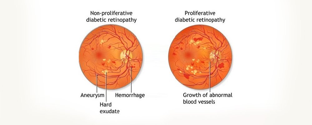
Diabetic retinopathy diagnosis and management – Screening for diabetic retinopathy is important for individuals having diabetes of five years or more duration. It can destroy the vision silently over a period of time. Patients if diagnosed would need FFA and/or OCT to define the stage of the disease. Advanced cases might need retinal laser treatment or vitrectomy surgery to restore the vision.
Hypertensive retinopathy – Hypertensive retinopathy can affect the eye quietly or can present as loss of vision with sudden onset. Patients with hypertension needs to be examined from time to time and if evidence of retinopathy is found, they would need FFA, OCT, retinal laser or vitrectomy (surgery) depending on the kind of damage.
Age related retinopathies – Crucial areas of the retina can get affected during the old age. Generally the disease affects the people with age more than 50. Vision loss is generally slow and progressive. In few unfortunate patients there can be sudden acceleration of the disease process as well. FFA and OCT are required to diagnose the extent of the disease. Treatment options include medications, intravitreal injections, lasers, telescopic iol implantation and vitrectomy depending on the extent of injury.
Retinal detachment management – Retinal detachment generally happens in people with minus power glasses. It can also happen secondary to trauma, complicated surgery, other vascular retinopathies, some genetic disorders, pediatric retinopathies etc. This is a surgical problem requiring scleral buckling or vitrectomy with/without gas or silicon oil injection.
Cataract complications management – Vision threatening complications of the cataract surgery like dropped nucleus, dropped IOL or endophthalmitis require retinal surgery to restore the vision. Our institute excels in scleral fixated IOL
Ocular trauma – Injuries of the eye affecting the inside portion with affected vision or threatened affected vision requires surgical exploration and restoration of the normal anatomy.
Pediatric retina – Do you see a white pearl your child’s eye? If yes, then contact us immediately. Most of the retinal disorders present in children like this. The condition can be as simple as bleeding at the back of the eye to serious disorders like tumours. Treatment depends on the problem child is facing. Premature child with low birth weight needs retinal evaluation early in the life to rule our ROP a vision threatening condition.
Congenital abnormailities & retinopathies – Patients suffering from these conditions are generally young. They need a through evaluation of the eye and need the eletrophysiological tests (VEP, ERG, EOG). Most of the conditions are managed conservatively.
Endophthalmitis management – The term represents a serious inflammation into the eye. Although there can be many causes leading to this problem most of the cases are secondary to some intraocular surgery. Patients require immediate intervention which may be in the form of multiple surgeries.
Investigations
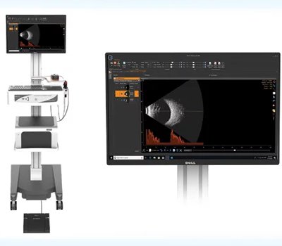
B scan – Is the ultrasound of the eye. It gives a two dimentional picture of the eye from various angles. The procedure is simple where a vibrating probe is kept over the eye. The whole procedure spans 5 minutes.
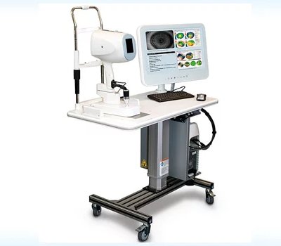
FFA – It is a 20 minute procedure. A green dye is injected into the arm and eye is photographed. The procedure is needed to determine the small subclinical problems with the blood supply of the retina. The test requires dilated pupil.
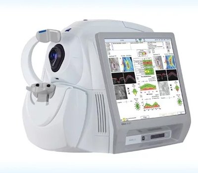
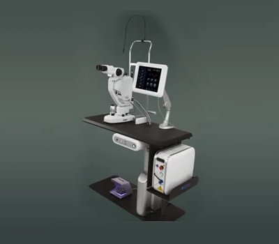
OCT – It is a 5 minute procedure. A laser beam is dirested into the eye to scan the crucial areas of the eye. The test determines the thickness of the retina and helps in highlighting the problems in various layers of the retina. The test requires dilated pupil.
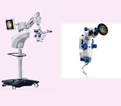
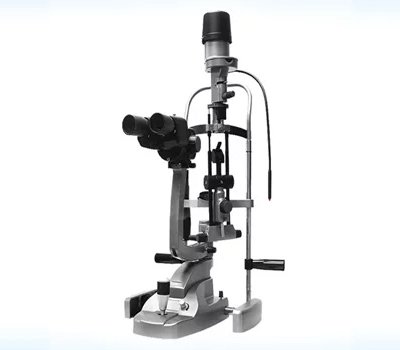
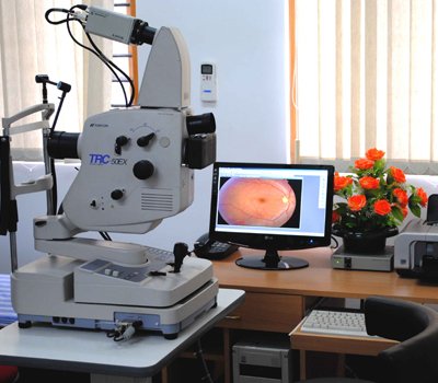
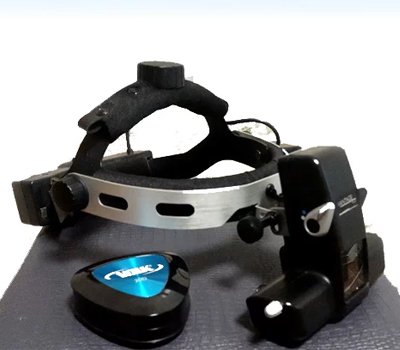
INDIRECT OPHTHALMOSCOPY – It is a simple test of 5 minutes duration. Retina is examined through a beam of light going into the eye. It is a basic test and requires dilated pupil.
Treatment Medical
Periocular injections – A simple injection given at the outermost layer of the eye ball to treat fluid collections at the crucial areas of the retina.
Intraocular injections – The injection is given inside the eye. The procedure is done under all aseptic conditions under topical (drop) anesthesia.
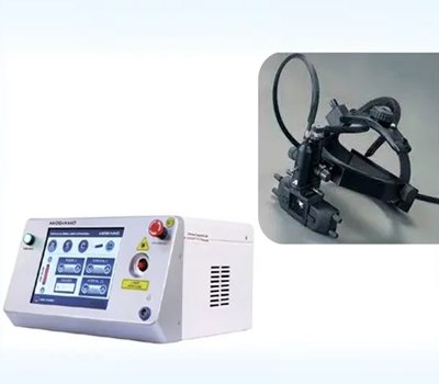
Retinal lasers – A 10 minute procedure done in OPD. Needs pupillary dilatation. Does not need any anesthesia. The defective areas on the retina are burned with the laser. Mainly used to treat vasular retinopathies
Laser for myopic retinopathies – A 10 minute procedure to delimit the retinal breaks to prevent a serious complication called retinal detachment. Needs pupillary dilatation. Does not need any anesthesia.
Yag lasers for after cataracts – A two minute no anesthesia procedure to clean the artificial lens implanted in the eye during an earlier cataract surgery.
Surgical
Vitrectomy 20/23/25/27 guage – conventional method to perform a retinal surgery. Three holes are created in the eye to visualise and operate the retinal problem. The surgery is done under local anesthesia. Minmum duration of the surgery is 30 minutes. The patient might be implanted with gas or oil inside the eye. The oil needs to be removed later on. It is very important to maintain the position suggested by the doctor after the surgery.
Scleral buckling – A classical method to treat retinal detachments where a band or buckle is implanted around the eye to support the slipping retina. Minimum duation of surgery is 40 minutes and is performed under local anesthesia. Cryotherapy for retinal lesions – A 10 minute procedure performed under local anaesthesia. Here the early lesions on the retina are freezed with a cryopexy probe to prevent retinal detachment.
Cyclodestructive procedure – Cryopexy probe is used to freeze the pressure maintaining tissue in the eye to control pressures in the eyes with uncontrolled gaucoma.
Traumatic globe repairs – Surgical procedure depends on the kind of presentation the patient has. The management can range from simple suturing to complicated combined procedures like combination of scleral buckling with vitrectomy.